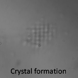
The technique could someday be used to analyze the structure of materials of biological interest, including bacteria, viruses and proteins, said U-M physicist Georg Raithel.
Raithel is co-author of a research paper on the topic published online May 31 in the journal Physical Review E. The other author is U-M research fellow Betty Slama-Eliau.
The standard method used to characterize biological molecules like proteins involves crystallizing them, then analyzing their structure by bombarding the crystals with X-rays, a technique called X-ray crystallography. But the method cannot be used on many of the proteins of highest interestâ€"such as cell-membrane proteinsâ€"because there's no way to crystallize those molecules.
"So we came up with this idea that one could use, instead of a conventional crystal, an optically induced crystal in order to get the crystallization of a sample that could be suitable for structural analysis," said Raithel, professor of physics and associate chair of the department.
 To move toward that goal, Raithel and his colleagues are developing the laser technique using microscopically small plastic spheres instead of the molecules. Other researchers have created 3-D optically induced crystals, but Raithel said the crystals his team created are denser than those previously achieved.
The process involves shining laser beams through two opposed microscope lenses, one directly beneath the other. Two infrared laser beams are directed through each lens, and they meet at a common focal point on a microscope slide that holds thousands of plastic nanoparticles suspended in a drop of water.
The intersecting laser beams create electric fields that vary in strength in a regular pattern that forms a 3-D grid called an optical lattice. The nanoparticles get sucked into regions of high electric-field strength, and thousands of them align to form optically induced crystals. The crystals are spherical in shape and about 5 microns in diameter. A micron is one millionth of a meter.
Imagine an egg crate containing hundreds of eggs. The cardboard structure of the crate is the optical lattice, and each of the eggs represents one of the nanoparticles. Stack several crates on top of each other and you get a 3-D crystal structure.
"The crate is the equivalent of the optical lattice that the laser beams make," Raithel said. "The structure of the crystal is determined by the egg carton, not by the eggs."
The optical crystals dissipate as soon as the laser is switched off.
The research was funded by the National Science Foundation.
Contact: Jim Erickson ericksn@umich.edu 734-647-1842 University of Michigan
